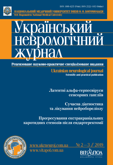
Вплив мікробіоти кишечника та моделі харчування на патогенез та перебіг розсіяного склерозу
DOI:
https://doi.org/10.30978/UNJ2019-2-22Ключові слова:
розсіяний склероз, кишкова мікробіота, мікробіом кишечника, автоімунні захворювання, дієтичний патернАнотація
Вивчення ролі кишкової мікробіоти у патогенезі автоімунних та демієлінізувальних захворювань має важливе значення. Отримані протягом останнього десятиліття експериментальні дані свідчать про тісний зв’язок між імунною системою та мікробіотою кишечника. У проаналізованій літературі знайдено додаткове підтвердження участі мікробіоти кишечника в імунорегуляції та розвитку імуноопосередкованих захворювань: ревматоїдного артриту, псоріазу, синдрому Шегрена, розсіяного склерозу, метаболічного синдрому, автоімунного гепатиту, цукрового діабету 1 типу, спондилоартриту, запальних захворювань кишечника. Дослідження останніх років продемонстрували, що кишкова мікробіота за допомогою численних мікробних сполучень, вегетативної нервової системи, продукування нейрометаболітів, сигнальних молекул та нейротрансмітерів чинить вплив на функціонування центральної нервової системи. Враховуючи це, запропоновано модель осі «кишечник — мозок», яка передбачає взаєморегуляцію між кишковою мікробіотою та центральною нервовою системою. Виявлення впливу кишкової мікробіоти на функціонування імунної та нервової систем зумовило підвищений науковий інтерес до мікробіоти кишечника як етіопатогенетичного фактора розвитку такого автоімунного захворювання, як розсіяний склероз. Виявлено, що склад та особливості кишкової мікробіоти у хворих на розсіяний склероз порівняно зі здоровими особами відрізняються. Поки що точно не встановлено, який тип мікроорганізмів асоційований з розвитком цього захворювання та характером його перебігу. Тому для пошуку способів корекції порушень у пацієнтів із розсіяним склерозом необхідним є вивчення складу кишкової мікробіоти. Важливою складовою таких досліджень є оцінка моделі харчування, оскільки саме дієтичний патерн визначає ентеротип людини та впливає на концентрацію та розподіл мікроорганізмів у кишечнику. Тому необхідно зіставляти результати дослідження складу кишкової мікробіоти з типом дієтичного патерну пацієнта.
Посилання
Podolinskaya NA, Vykhrystsenka LR. The role of the intestinal microflora in the regulation of the immune response in rheumatoid arthritis. Immunopathology, allergology, infectology (Russian). 2017;1:37-45. doi: 10.14427/jipai.2017.1.37.
Starovoitova SO, Karpov OV. Prospects for the use of probiotic microorganisms in functional foods and medicine. Food industry (Ukrainian). 2015;18:76-80.
Tkach SM et al. Intestinal microbiota and functional diseases of the intestine,. Modern Gastroenterology (Russian). 2014;1 (75):116-119.
Alkanani, Aimon K et al. Alterations in Intestinal Microbiota Correlate With Susceptibility to Type 1 Diabetes. Diabetes Vol 64,10. 2015:3510-3520. doi:10.2337/db14-1847.
Amini M, Esmaillzadeh A, Shafaeizadeh S, Behrooz J, Zare M. Relationship between major dietary patterns and metabolic syndrome among individuals with impaired glucose tolerance. Nutrition. 2010;26 (10):986-992. doi: 10.1016/j.nut.2010.03.006. Epub 2010 Jul 10.
Atarashi K, Tanoue T, Shima T et al. Induction of colonic regulatory T cells by indigenous Clostridium species. Science. 2011;331 (6015):337-341. Epub 2010 Dec 23. https://doi.org/10.1126/science.1198469.
Atlas of Multiple sclerosis, Multiple Sclerosis International Federation, 2013.
Bailey MT, Dowd SE, Galley JD, Hufnagle AR, Allen RG, Lyte M. Exposure to a social stressor alters the structure of the intestinal microbiota: implications for stressor-induced immunomodulation. Brain, Behavior, and Immunity. 2011;25 (3):397-407. doi: 10.1016/j.bbi.2010.10.023.
Barrett E, Ross RP, O’Toole PW, Fitzgerald GF, Stanton C. γ-Aminobutyric acid production by culturable bacteria from the human intestine. J Appl Microbiol. 2012;113 (2). 411-417. doi: 10.1111/j.1365-2672.2012.05344.x.
Bellavance MA, Rivest S. The HPA — immune axis and the immunomodulatory actions of glucocorticoids in the brain. Frontiers in Immunology. 2014;5:136. doi: 10.3389/fimmu.2014.00136.
Branton WG, Lu JQ, Surette MG et al. Brain microbiota disruption within inflammatory demyelinating lesions in multiple sclerosis. Scientific Reports. 2016;6 (1, article 37344) doi: 10.1038/srep37344.
Chen J, Chia N, Kalari KR et al. Multiple sclerosis patients have a distinct gut microbiota compared to healthy controls. Scientific Reports. 2016;6 (1, article 28484) doi: 10.1038/srep28484.
Chu F, Shi M, Lang Y et al. Gut microbiota in multiple sclerosis and experimental autoimmune encephalomyelitis: current applications and future perspectives. Mediators Inflamm. 2018;2018:8168717. doi: 10.1155/2018/8168717. PubMed PMID: 29805314. PubMed Central PMCID: PMC5902007.
De Luca F, Shoenfeld Y. The microbiome in autoimmune diseases. Clin Exp Immunol. 2018;195:74-85.
Fleck AK, Schuppan D, Wiendl H, Klotz L. Gut-CNS-Axis as possibility to modulate inflammatory disease activity-implications for multiple sclerosis. Int J Mol Sci. 2017;18 (7):1526.
Furusawa Y, Obata Y, Fukuda S et al. Commensal microbe-derived butyrate induces the differentiation of colonic regulatory T cells. Nature. 2013;504 (7480):446-450. doi: 10.1038/nature12721.
Gill T, Asquith M, Rosenbaum JT, Colbert RA. The intestinal microbiome in spondyloarthritis. Curr Opin Rheumatol. 2015;27 (4):319-325. doi:10.1097/BOR.0000000000000187.
Giongo, A. et al. Toward defining the autoimmune microbiome for type 1 diabetes. ISME J. 2011;5:82-91.
Hugon P, Dufour JC, Colson P, Fournier PE, Sallah K, Raoult D. A comprehensive repertoire of prokaryotic species identified in human beings. Lancet. Infectious Diseases. 2015;15 (10):1211-1219. doi: 10.1016/S1473-3099 (15)00293-5.
Human Microbiome Project C. Structure, function and diversity of the healthy human microbiome. Nature. 2012;486:207-214. PMID:22699609. https://doi.org/10.1038/nature11234.
Jahromi SR, Toghae M, Jahromi MJ, Aloosh M. Dietary pattern and risk of multiple sclerosis. Iran J Neurol. 2012;11 (2):47-53. PubMed PMID: 24250861. PubMed Central PMCID: PMC3829243.
Jangi S, Gandhi R, Cox LM et al. Alterations of the human gut microbiome in multiple sclerosis. Nature Communications. 2016;7, article 12015 doi: 10.1038/ncomms12015.
Kamdar K, Nguyen V, DePaolo R. Toll-like receptorsignaling and regulation of intestinal immunity. Virulence. 2013;4, N3 P.207-212.
Kirby T, Ochoa-Repáraz J. The Gut Microbiome in Multiple Sclerosis: A Potential Therapeutic Avenue. Med Sci (Basel). 2018;6 (3):69. doi: 10.3390/medsci6030069.
Li J, Jia H, Cai X et al. An integrated catalog of reference genes in the human gut microbiome. Nature Biotechnology. 2014;32 (8):834-841. doi: 10.1038/nbt.2942.
Lin R et al. Abnormal intestinal permeability and microbiota in patients with autoimmune hepatitis. Int J Clin Exp Pathol. 2015;8:5153-5160.
Louis P, Flint HJ. Formation of propionate and butyrate by the human colonic microbiota. Environmental Microbiology. 2017;19 (1):29-41. doi: 10.1111/1462-2920.13589.
Lyte M. Microbial endocrinology in the microbiome-gut-brain axis: how bacterial production and utilization of neurochemicals influence behavior. PLoS Pathogens. 2013;9 (11, article e1003726) doi: 10.1371/journal.ppat.1003726.
Margherita T. Vitamin D and its role in immunology: multiple sclerosis, and inflammatory bowel disease. Prog Biophys Mol Biol. 2006;92(1):60-64.
Mayer EA, Tillisch K, Gupta A. Gut/brain axis and the microbiota. J Clin Invest. 2015;125 (3):926-938. doi: 10.1172/JCI76304.
Okun, T., Kinoshita M, Ishikura T, Mochizuki H. Role of diet, gut microbiota, and metabolism in multiple sclerosis and neuromyelitis optica. Clin Exp Neuroimmunol. 2019;10:12-19. doi:10.1111/cen3.12499.
Round JL, Mazmanian SK. Inducible Foxp3+ regulatory T-cell development by a commensal bacterium of the intestinal microbiota. Proc Natl Acad Sci USA. 2010;107:12204-12209. doi: 10.1073/pnas.0909122107.
Sachiko M, Sangwan K, Wataru S et al. Dysbiosis in the Gut Microbiota of Patients with Multiple Sclerosis, with a Striking Depletion of Species Belonging to Clostridia XIVa and IV Clusters. PLoS One. 2015;10 (9). 0137429.
Salem I, Ramser A, Isham N, Ghannoum MA. The gut microbiome as a major regulator of the gut-skin axis. Frontiers in Microbiology. 2018;9. 1459. doi:10.3389/fmicb.2018.01459.
Schirmer M, Smeekens SP, Vlamakis H et al. Linking the Human Gut Microbiome to Inflammatory Cytokine Production Capacity. Cell. 2016;167 (4):1125-1136.e8. doi:10.1016/j.cell.2016.10.020.
Shahi SK, Freedman SN, Mangalam AK. Gut microbiome in multiple sclerosis: The players involved and the roles they play. Gut Microbes. 2017;8 (6):607-615. doi:10.1080/19490976.2017.1349041.
Steimle A, Frick J:Molecular Mechanisms of Induction of Tolerant and Tolerogenic Intestinal Dendritic Cells in Mice. J Immunol Res. 2016;1958650. Published online 2016 Feb 11. https://doi.org/10.1155/2016/1958650.
Tourtas T, Birke MT, Kruse FE, Welge-Lüssen UC, Birke K. Preventive effects of omega-3 and omega-6 Fatty acids on peroxide mediated oxidative stress responses in primary human trabecular meshwork cells. PloS One. 2012;7 (2):e31340. doi:10.1371/journal.pone.0031340.
Tsigalou C, Stavropoulou E, Bezirtzoglou E. Current Insights in Microbiome Shifts in Sjogren’s Syndrome and Possible Therapeutic Interventions. Front Immunol. 2018;9:1106. doi:10.3389/fimmu.2018.01106.
Umeton R, Eleftheriou E, Nedelcu S et al. The gut microbiome in relapsing multiple sclerosis patients compared to controls. Neurology. 2018;90 (suppl.. 15). P2.355.
Warren S, Warren KG. Multiple sclerosis. World Health Organization, Geneva, 2001.


