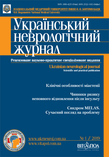
Структурні зміни головного мозку на етапі клінічно ізольованого розсіяного склерозу
DOI:
https://doi.org/10.30978/UNJ2019-1-26Ключові слова:
клінічно ізольований синдром, розсіяний склероз, атрофіяАнотація
Мета — визначити обсяг атрофії головного мозку в пацієнтів із клінічно ізольованим синдромом (КІС) порівняно зі здоровими особами.
Матеріали і методи. Обсерваційне поперечне контрольоване дослідження атрофічних змін головного мозку в пацієнтів з КІС розсіяного склерозу проведене вперше в Україні. Обстежено 30 осіб з КІС. Контрольну групу утворили 29 здорових осіб. Середній вік пацієнтів з КІС становив (32,07 ± 8,54) року, здорових осіб — (36,70 ± 2,39) року (р = 0,02). В обох групах переважали жінки — відповідно 27 (90 %) і 18 (62 %) (р = 0,07). У 18 (60 %) пацієнтів виявлено мультифокальний КІС, у решти — монофокальний КІС. Кількість балів за функціональними шкалами EDSS була такою: очна — (0,833 ± 1,090) бала, пірамідна — (1,33 ± 0,96) бала, мозочкова — (1,33 ± 1,12) бала, тазові розлади — (0,23 ± 0,63) бала, церебральні порушення — (0,17 ± 0,38) бала. Загальний бал за шкалою EDSS на момент КІС — (2,98 ± 0,55) бала. Для оцінки атрофічних процесів у всіх пацієнтів та здорових осіб вимірювали 23 лінійних параметри та обчислювали 14 індексів для кожної магнітно‑резонансної томограми.
Результати. Рання атрофія, виявлена в пацієнтів з КІС порівняно з контрольною групою, відбувається переважно в сірій речовині головного мозку. Статистично значущу (p < 0,05) різницю між групами встановлено за індексом серединних структур головного мозку, шириною третього та бічних шлуночків. Індекси мозолистого тіла, фронтальної атрофії, Еванса, Гукмана, бікаудатний і шлуночковий парієто‑окципітальний є недостатньо чутливими для оцінки атрофічних змін головного мозку у хворих з КІС. Механізм нейродегенерації запускається при КІС на початку захворювання, ймовірно, раніше, ніж клінічний початок захворювання.
Висновки. Встановлено, що атрофічні процеси головного мозку відбуваються на етапі КІС. Індекс серединних структур головного мозку, ширина третього та бічних шлуночків є найчутливішими показниками для оцінки атрофії мозку на етапі КІС.
Посилання
Calabrese M, Rinaldi F, Mattisi I et al. The predictive value of gray matter atrophy in clinically isolated syndromes. Neurology. 2011;77(3):257-263. PubMed PMID:21613600.
Ceccarelli A, Rocca MA, Pagani E et al. A voxelbased morphometry study of grey matter loss in MS patients with different clinical phenotypes. NeuroImage. 2008;42(1):315-322. PubMed PMID: 18501636.
Ceccarelli A, Rocca MA, Perego E et al. Deep grey matter T2 hypo-intensity in patients with paediatric multiple sclerosis. Multiple sclerosis. 2011;17(6):702-707. PubMed PMID: 21228024.
De Stefano N, Sprenger T, Freedman MS et al. Including threshold rates of brain volume loss in the definition of disease activity-free in multiple sclerosis using fingolimod phase 3 data. Proceedings of Joint ACTRIMS-ECTRIMS Meeting Boston, MA, US 10-13 September 2014. Moda access : http://www.abstractstosubmit.com/msboston2014/eposter/main.php? do¼YToyOntzOjU6Im1vZHVsIjtzOjY6ImRldGFpbCI7czo4OiJkb2N1bWVudCI7a To3NzQ7fQ¼ ¼&
De Stefano N, Stromillo ML, Giorgio A et al. Establishing pathological cut-offs of brain atrophy rates in multiple sclerosis. J Neurol Neurosurg Psychiatry. 2016;87:93-99. Doi: 10.1136/jnnp-2014-309903
Figueira F, Santos V, Figueira G, Silva A. A practical method for long-term follow-up in multiple sclerosis. ArqNeuropsiquiatr. 2007;65 (4-A):931-935.
Giovannoni G. et al. Brain health: time matters in multiple sclerosis. Multiple Sclerosis and Related Disorders. 2016;9:S5-S48. Doi: org/10.1016/j.msard.2016.07.003
Hagemeier J, Weinstock-Guttman B, Bergsland N et al. The use of corpus callosum index in the measurement of brain atrophy in multiple sclerosis. Egypt J Neurol Psychiat Neurosurg. 2010;47(4):633-637.
Henry RG, Shieh M, Okuda DT et al. Regional grey matter atrophy in clinically isolated syndromes at presentation. Journal of Neurology, Neurosurgery & Psychiatry. 2008;79:1236-1244.
Kennedy C et al. Iron deposition on SWI-filtered phase in the subcortical deep gray matter of patients with clinically isolated syndrome may precede structure-specific atrophy. Am J neuroradiol. 2012 Sep;33(8):1596-601. PubMed PMID: 22460343.
Kizlaitienandedot R, Kaubrys G, Giedraitienandedot N, Ramanauskas N. Dementaviandccaron; ienandedot; J. Composite marker of cognitive dysfunction and brain atrophy is highly accurate in discriminating between relapsing-remitting and secondary progressive multiple sclerosis. medical science monitor. International Medical Journal of Experimental and Clinical Research. 2017;23:588-597. Doi: 10.12659/MSM.903234.
Kurtzke JF. Rating Neurologic Impairment in Multiple Sclerosis an Expanded Disability Status Scale (EDSS). Neurology. 1983;33:1444.
Menendez M, Arias-Carrión O. Indices of regional brain atrophy: Formulae and Nomenclature. Cureus. 2015;7(8):e295. Doi: 10.7759/cureus.295.
Miller DH, Chard DT, Ciccarelli O. Clinically isolated syndromes. Lancet Neurol. 2012;11:157-169. [PubMed]
Missori P, Currà A. Progressive cognitive impairment evolving to dementia parallels parieto-occipital and temporal enlargement in idiopathic chronic hydrocephalus: A retrospective cohort study. Frontiers in Neurology. 2015;6:15. Doi: 10.3389/fneur.2015.00015.
Montalban X, Gold R, Thompson J et al. ECTRIMS/EAN Guideline on the pharmacological treatment of people with multiple sclerosis. European Journal of Neurology. 2018;25. 10.1111/ene.13536.
Multiple Sclerosis International Federation, 2013. Atlas of MS Database Data Export: Number of People with MS, 2013. Multiple Sclerosis International Federation. Moda access: http://www.atlasofms.org.
Naud A, Schmitt E, Wirth M, Hascoet J. M. Determinants of indices of cerebral volume in former very premature infants at term equivalent age. PLoS ONE. 2017;12(1):e0170797. Doi: 10.1371/journal.pone.0170797.
Polman CH, Reingold SC, Banwell B et al. Diagnostic criteria for multiple sclerosis: 2010 revisions to the McDonald criteria. Annals of Neurology. 2011;69(2):292-302. Doi: 10.1002/ana.22366
Tompson AJ et al. Diagnosis of multiple sclerosis: 2017 revisions of the McDonald criteria. Lancet Neurology. 2017:162-173.
Tsivgoulis G, Katsanos AH, Grigoriadis N et al. The effect of disease-modifying therapies on brain atrophy in patients with clinically isolated syndrome: a systematic review and meta-analysis. Ther Adv Neurol Disord. 2015;8(5):193-202.
Zivadinov R, Havrdová E, Bergsland N et al. Thalamic atrophy is associated with development of clinically definite multiple sclerosis. Radiology. 2013;268(3):831-841. PubMed PMID: 23613615.


