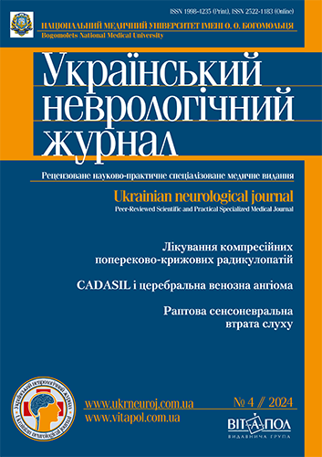
CADASIL і церебральна венозна ангіома. Клінічний випадок та огляд літератури
DOI:
https://doi.org/10.30978/UNJ2024-4-51Ключові слова:
CADASIL; церебральна автосомно-домінантна артеріопатія; підкіркові інфаркти; лейкоенцефалопатія; NOTCH3; церебральна венозна ангіома; перицит; підкіркова судинна деменція.Анотація
Представлено огляд наукової літератури та опис клінічного випадку поєднаної артеріальної та венозної цереброваскулярної патології — церебральної автосомно-домінантної артеріопатії з підкірковими інфарктами й лейкоенцефалопатією (CADASIL) і венозної ангіоми мозку. CADASIL належить до рідкісних захворювань нервової системи, але водночас є найпоширенішою моногенною ангіопатією головного мозку, що призводить до раннього виникнення гострих і хронічних розладів мозкового кровообігу. Висвітлено сучасні погляди на епідеміологію, етіопатогенез, клініку, діагностику та менеджмент зазначеної патології. Представлені оновлені дані щодо поширеності типових і атипових мутацій гена NOTCH3, значення накопичення в судинній стінці змінених білків для розвитку селективної вазоконстрикції та інших етапів каскаду патологічних судинних порушень, а також щодо ролі перицитів у розвитку генетично детермінованої васкулопатії. Наведено результати досліджень корелятивних зв’язків між варіантами мутацій гена NOTCH3 та клінічною картиною захворювання, а також аналіз особливостей генотипу й фенотипу в пацієнтів із CADASIL у різних регіонах. Проведено ретроспективний аналіз становлення сучасних клінічних критеріїв CADASIL, які мають високу чутливість і специфічність та забезпечують швидкий скринінговий відбір пацієнтів для генетичного тестування з метою остаточної верифікації діагнозу. Використання клінічних критеріїв як діагностичного інструмента показано на прикладі власного клінічного спостереження. Описано еволюцію терапевтичних підходів і вторинної профілактики судинних уражень великого мозку за наявності CADASIL.
Посилання
Dovbonos TA. [Tserebralna avtosomno-dominantna arteriopatiia iz subkortykalnymy infarktamy i leikoentsefalopatiieiu (CADASIL)]. Ukrainian Neurological Journal. 2013;1:22-8. Ukrainian.
Prokopiv MM, Orel MIa, Vakulenko LO, Kovalenko NV. [Klinichnyi vypadok syndromu CADASIL]. Ukrainskyi medychnyi chasopys. 2024;(5)IX/X. http://doi.org/10.32471/umj.1680-3051.163.252316. Ukrainian.
Aghetti A, Amsellem T, Hervé D, Chabriat H, Guey S. Border-zone cerebral infarcts associated with COVID-19 in CADASIL: A report of 3 cases and literature review. Cerebrovasc Dis Extra. 2024;14(1):1-8. http://doi.org/10.1159/000534975.
Aguilar-Fuentes V, Justo-Hernandez D, Arredondo-Dubois JM, et al. Palliative care in CADASIL: diagnosis is only the first step. Arq Neurosiquiatr. 2023;81(11):1022-4. http://doi.org/10.1055/s-0043-1777009.
Alqarni AA, Shirah B, Algahtani H, Almohiy H, Hassan A. Cerebral autosomal dominant arteriopathy with subcortical infarcts and leukoencephalopathy: Atypical clinical presentation with isolated frontotemporal dementia. Journal of Neurosciences in Rural Practice. 2023 Apr-Jun;14(2):371-3. http://doi.org/10.25259/jnrp_88_2023.
Ameer MA, Bhutta BS, Asghar N, Haseeb MT, Abbasi RN. Cerebral Autosomal Dominant Arteriopathy with Subcortical Infarcts and Leukoencephalopathy (CADASIL) Presenting as Migraine. Cureus. 2021 May;13(5):e15355. http://doi.org/10.7759/cureus.15355.
Ashraf S, Allena N, Shrestha E, Dhallu M, Khaja M. Cerebral Autosomal Dominant Arteriopathy with Subcortical Infarcts and Leukoencephalopathy (CADASIL): A rare cause of transient ischemic attack. Cureus. 2022 Oct;14(10):e30940. http://doi.org/10.7759/cureus.30940.
Benedetti V, Canzoneri R, Perrelli A, et al. Next-generation sequencing advances the genetic diagnosis of Cerebral Cavernous Malformation (CCM). Antioxidants (Basel). 2022 Jun 29;11(7):1294. http://doi.org/10.3390/antiox11071294.
Berge E, Whiteley W, Audebert H, De Marchis GM, Fonseca AC, Padiglioni C, de la Ossa NP, Strbian D, Tsivgoulis G, Turc G. European Stroke Organisation (ESO) guidelines on intravenous thrombolysis for acute ischaemic stroke. Eur Stroke J. 2021;6:I-LXII. http://doi.org/10.1177/2396987321989865.
Cao Y, Zhang D-D, Han F, Jiang N, Yao M, Zhu Y-C. Phenotypes associated with NOTCH3 cysteine-sparing mutations in patients with clinical suspicion of CADASIL: a systematic review. Int J Mol Sci. 2024;25:8796. http://doi.org/10.3390/ijms25168796.
Chojdak-Łukasiewicz J, Dziadkowiak E, Budrewicz S. Monogenic causes of strokes. Genes (Basel). 2021 Nov 23;12(12):1855. http://doi.org/10.3390/genes12121855.
Cruciani A, Pilato F, Rossi M, Motolese F, Di Lazzaro V, Capone F. Ischemic stroke in a patient with stable CADASIL during COVID-19: a case report. Brain Sci. 2021 Dec 8;11(12):1615. http://doi.org/10.3390/brainsci11121615.
D’Souza D, Sharma R, Ranchod A, et al. Cerebral autosomal dominant arteriopathy with subcortical infarcts and leukoencephalopathy (CADASIL). Reference article, Radiopaedia.org (Accessed on 11 Apr 2024). http://doi.org/10.53347/rID-1027.
Dai Z, Li J, Li Y, Wang R, Yan H, et al. Role of pericytes in the development of cerebral cavernous malformations. iScience. 2022;25(12):105642. http://doi.org/10.1016/j.isci.2022.105642.
Davous P. CADASIL: a review with proposed diagnostic criteria. Eur J Neurol. 1998 May;5(3):219-33. http://doi.org/10.1046/j.1468-1331.1998.530219.x.
Di Donato I, Bianchi S, De Stefano N, et al. Cerebral Autosomal Dominant Arteriopathy with Subcortical Infarcts and Leukoencephalopathy (CADASIL) as a model of small vessel disease: update on clinical, diagnostic, and management aspects. BMC Med. 2017;15(1):41. http://doi.org/10.1186/s12916-017-0778-8.
Ding R, Hase Y, Ameen-Ali KE, et al. Loss of capillary pericytes and the blood-brain barrier in white matter in poststroke and vascular dementias and Alzheimer’s disease. Brain Pathol. 2020;30:1087-101. http://doi.org/10.1111/bpa.12888.
Dupé C, Guey S, Biard L, et al. Phenotypic variability in 446 CADASIL patients: Impact of NOTCH3 gene mutation location in addition to the effects of age, sex, and vascular risk factors. J Cereb Blood Flow Metab. 2023;43:153-66. http://doi.org/10.1177/0271678X221126280.
Fiebig A, Krusche P, Wolf A, et al. Heritability of chronic venous disease. Hum Genet. 2010 Jun;127(6):669-74. http://doi.org/10.1007/s00439-010-0812-9.
Glover PA, Goldstein ED, Badi MK, et al. Treatment of migraine in patients with CADASIL: A systematic review and meta-analysis. Neurol Clin Pract. 2020 Dec;10(6):488-96. http://doi.org/10.1212/CPJ.0000000000000769.
Goh JW, Kundu S, Durairajan R. Cerebral Autosomal Dominant Arteriopathy with Subcortical Infarcts and Leukoencephalopathy (CADASIL): A diagnosis to consider in atypical stroke presentations. Cureus. 2023 Oct;15(10):e46482. http://doi.org/10.7759/cureus.46482.
Guey S, Mawet J, Hervé D, et al. Prevalence and characteristics of migraine in CADASIL. Cephalalgia. 2016 Oct;36(11):1038-47. http://doi.org/10.1177/0333102415620909.
Gürler G, Soylu KO, Yemisci M. Importance of pericytes in the pathophysiology of cerebral ischemia. Noro Psikiyatr Ars. 2022 Dec 16;59(Suppl 1):S29-S35. PMID: 36578988; PMCID: PMC9767130.
Hack RJ, Cerfontaine MN, Gravesteijn G, et al. Effect of NOTCH3 EGFr Group, Sex, and Cardiovascular Risk Factors on CADASIL Clinical and Neuroimaging Outcomes. Stroke. 2022;53:3133-44. http://doi.org/10.1161/STROKEAHA.122.039325.
Huang H, Xie W, Hu F, Lv H, Wu Y, Cai B. Acute bilateral multiple subcortical infarcts as a manifestation in cerebral autosomal dominant arteriopathy with subcortical infarcts and leukoencephalopathy. Neurol Sci. 2023 Dec;44(12):4391-9. http://doi.org/10.1007/s10072-023-06949-9.
Jiménez-Sánchez L, Hamilton OKL, Clancy U, et al. Sex differences in cerebral small vessel disease: a systematic review and Meta-analysis. Front Neurol. 2021;12:12. http://doi.org/10.3389/fneur.2021.756887.
Karim R, Malik M, Cheema H, Aziz A, Khan R. Cerebral Autosomal Dominant Arteriopathy with Subcortical Infarcts and Leukoencephalopathy (CADASIL) in a 32-year-old male presenting with a Transient Ischemic Attack (TIA). Cureus. 2024 Oct;16(10):e70970. http://doi.org/10.7759/cureus.70970.
Kim H, Lim YM, Lee EJ, Oh YJ, Kim KK. Clinical and imaging features of patients with cerebral autosomal dominant arteriopathy with subcortical infarcts and leukoencephalopathy and cysteine-sparing NOTCH3 mutations. PLoS One. 2020;15:e0234797. http://doi.org/10.1371/journal.pone.0234797.
Liao YC, Hu YC, Chung CP, et al. Intracerebral hemorrhage in cerebral autosomal dominant arteriopathy with subcortical infarcts and leukoencephalopathy: prevalence, clinical and neuroimaging features and risk factors. Stroke. 2021 Mar;52(3):985-93. http://doi.org/10.1161/STROKEAHA.120.030664.
Lin H-J, Chen C-H, Su M-W, et al. Modifiable vascular risk factors contribute to stroke in 1080 NOTCH3 R544C carriers in Taiwan Biobank. Int J Stroke. 2024;19:105-13. http://doi.org/10.1177/17474930231191991.
Locatelli M, Padovani A, Pezzini A. Pathophysiological mechanisms and potential therapeutic targets in Cerebral Autosomal Dominant Arteriopathy with Subcortical Infarcts and Leukoencephalopathy (CADASIL). Front Pharmacol. 2020 Mar 13;11:321. http://doi.org/10.3389/fphar.2020.00321. PMID: 32231578; PMCID: PMC7082755.
Mancuso M, Arnold M, Bersano A, Burlina A, Chabriat H, Debette S, et al. Monogenic cerebral small-vessel diseases: diagnosis and therapy. Consensus recommendations of the European Academy of Neurology. Eur J Neurol. 2020;27:909-27. http://doi.org/10.1111/ene.14183.
Manini A, Pantoni L. CADASIL from bench to bedside: disease models and novel therapeutic approaches. Mol Neurobiol. 2021;58:2558-73. http://doi.org/10.1007/s12035-021-02282-4.
Manini A, Pantoni L. Genetic causes of cerebral small vessel diseases: a practical guide for neurologists. Neurology. 2023;100:766-83. http://doi.org/10.1212/WNL.0000000000201720.
Min J-Y, Park S-J, Kang E-J, et al. Mutation spectrum and genotype-phenotype correlations in 157 Korean CADASIL patients: a multicenter study. Neurogenetics. 2022;23:45-58. http://doi.org/10.1007/s10048-021-00674-1.
Mizuno T, Mizuta I, Watanabe-Hosomi A, Mukai M, Koizumi T. Clinical and genetic aspects of CADASIL. Front Aging Neurosci. 2020;12:91. http://doi.org/10.3389/fnagi.2020.00091.
Mizuta I, Nakao-Azuma Y, Yoshida H, Yamaguchi M, Mizuno T. Progress to clarify how NOTCH3 mutations lead to CADASIL, a hereditary cerebral small vessel disease. Biomolecules. 2024 Jan 18;14(1):127. http://doi.org/10.3390/biom14010127. PMID: 38254727; PMCID: PMC10813265.
Mizuta I, Watanabe-Hosomi A, Koizumi T, et al. New diagnostic criteria for cerebral autosomal dominant arteriopathy with subcortical infarcts and leukoencephalopathy in Japan. J Neurol Sci. 2017 Oct 15;381:62-7. http://doi.org/10.1016/j.jns.2017.08.009.
Muiño E, Fernández-Cadenas I, Arboix A. Contribution of «omic» studies to the understanding of Cadasil. A systematic review. Int J Mol Sci. 2021;22:7357. http://doi.org/10.3390/ijms22147357.
Muppa J, Yaghi S, Goldstein ED. Antiplatelet use and CADASIL: a retrospective observational analysis. Neurol Sci. 2023;44(8):2831-4. http://doi.org/10.1007/s10072-023-06773-1.
Ni W, Zhang Y, Zhang L, Xie JJ, Li HF, Wu ZY. Genetic spectrum of NOTCH3 and clinical phenotype of CADASIL patients in different populations. CNS Neurosci Ther. 2022;28(11):1779-89. http://doi.org/10.1111/cns.13917.
Pan AP, Potter T, Bako A, et al. Lifelong cerebrovascular disease burden among CADASIL patients: analysis from a global health research network. Front Neurol. 2023 Jul 14;14:1203985. http://doi.org/10.3389/fneur.2023.1203985. PMID: 37521283; PMCID: PMC10375407.
Paraskevas GP, Stefanou MI, Constantinides VC, et al. CADASIL in Greece: Mutational spectrum and clinical characteristics based on a systematic review and pooled analysis of published cases. Eur J Neurol. 2022;29:810-9. http://doi.org/10.1111/ene.15180.
Pescini F, Nannucci S, Bertaccini B,et al. The Cerebral Autosomal-Dominant Arteriopathy with Subcortical Infarcts and Leukoencephalopathy (CADASIL) Scale: a screening tool to select patients for NOTCH3 gene analysis. Stroke. 2012 Nov;43(11):2871-6. http://doi.org/10.1161/STROKEAHA.112.665927.
Pescini F, Torricelli S, Squitieri M, et al. Intravenous thrombolysis in CADASIL: report of two cases and a systematic review. Neurol Sci. 2023 Feb;44(2):491-8. http://doi.org/10.1007/s10072-022-06449-2.
Ruchoux MM, Kalaria RN, Román GC. The pericyte: A critical cell in the pathogenesis of CADASIL. Cerebral Circulation - Cognition and Behavior. 2021;2. https://doi.org/10.1016/j.cccb.2021.100031.
Rutten JW, van Eijsden BJ, Duering M, et al. The effect of NOTCH3 pathogenic variant position on CADASIL disease severity: NOTCH3 EGFr 1-6 pathogenic variants are associated with a more severe phenotype and lower survival compared with EGFr 7-34 pathogenic variant. Genet Med. 2019;21:676-82. http://doi.org/10.1038/s41436-018-0088-3.
Seyedaghamiri F, Geranmayeh MH, Ghadiri T, Ebrahimi-Kalan A, Hosseini L. A new insight into the role of pericytes in ischemic stroke. Acta Neurol Belg. 2023 Oct 7. http://doi.org/10.1007/s13760-023-02391-y. Epub ahead of print. PMID: 37805645.
Sukhonpanich N, Koohi F, Jolly AA, et al. Changes in the prognosis of CADASIL over time: a 23-year study in 555 individuals (2024) Journal of Neurology, Neurosurgery & Psychiatry Published Online First: 15 November. http://doi.org/10.1136/jnnp-2024-334823.
Sukhonpanich N, Markus HS. Prevalence, clinical characteristics, and risk factors of intracerebral haemorrhage in CADASIL: a case series and systematic review. J Neurol. 2024 May;271(5):2423-33. http://doi.org/10.1007/s00415-023-12177-0.
Wang J, Zhang L, Wu G, et al. Correction of a CADASIL point mutation using adenine base editors in hiPSCs and blood vessel organoids. J Genet Genomics. 2024 Feb;51(2):197-207. http://doi.org/10.1016/j.jgg.2023.04.013.
Williams OH, Mohideen S, Sen A, et al. Multiple internal border zone infarcts in a patient with COVID-19 and CADASIL. J Neurol Sci. 2020 Sep 15;416:116980. http://doi.org/10.1016/j.jns.2020.116980. Epub 2020 Jun 9. PMID: 32574902; PMCID: PMC7280138.
Winkler EA, Birk H, Burkhardt JK, et al. Reductions in brain pericytes are associated with arteriovenous malformation vascular instability. J Neurosurg. 2018 Dec 1;129(6):1464-74. http://doi.org/10.3171/2017.6.JNS17860. Epub 2018 Jan 5. PMID: 29303444; PMCID: PMC6033689.
Xiao S, Ke M, Cai K, Xu A, Chen M. Treatment options for patients with CADASIL and large-scale cerebral infarction: mechanical thrombectomy and antiplatelet therapy-A case report. Frontiers in Neurology. 2024;15:1400537. http://doi.org/10.3389/fneur.2024.1400537.
Yamamoto Y, Liao YC, Lee YC, Ihara M, Choi JC. Update on the epidemiology, pathogenesis, and biomarkers of cerebral autosomal dominant arteriopathy with subcortical infarcts and leukoencephalopathy. J Clin Neurol. 2023 Jan;19(1):12-27. http://doi.org/10.3988/jcn.2023.19.1.12.
Yan X, Shang J, Wang R, Wang F, Zhang J. Mechanisms regulating cerebral hypoperfusion in cerebral autosomal dominant arteriopathy with subcortical infarcts and leukoencephalopathy. Journal of Biomedical Research. 2022 Aug;36(5):353-7. http://doi.org/10.7555/jbr.36.20220208.
Yuan L, Chen X, Jankovic J, Deng H. CADASIL: A NOTCH3-associated cerebral small vessel disease. J Adv Res. 2024:S2090-1232(24)00001-8.
Zhang K, Loong SSE, Yuen LZH, et al. Genetics in ischemic stroke: current perspectives and future directions. J Cardiovasc Dev Dis. 2023 Dec 13;10(12):495. http://doi.org/10.3390/jcdd10120495. PMID: 38132662; PMCID: PMC10743455.
Zhang L, Zhang H, Zhou X, Zhao J, Wang X. Bibliometric analysis of research on migraine-stroke association from 2013 to 2023. J Pain Res. 2023 Dec 1;16:4089-112. http://doi.org/10.2147/JPR.S438745.
Zhang R, Ouin E, Grosset L, et al. Elderly CADASIL patients with intact neurological status. J Stroke. 2022;24(3):352-62. http://doi.org/10.5853/jos.2022.01578.
Zitser-Koren J, Crossland D, Driscoll T, et al. Characterization of a large single-site USA CADASIL cohort (2437). Neurology. 2021;96:2437. http://doi.org/10.1212/WNL.96.15_supplement.2437.
Zurrú MC, Casas Parera I, Moya G, Giovanelli C, Genovese O, Gatto E. CADASIL: un caso con diagnóstico molecular [Cadasil: a case with molecular diagnosis]. Medicina (B Aires). 2002;62(1):48-52. Spanish. PMID: 11965850.
##submission.downloads##
Опубліковано
Номер
Розділ
Ліцензія
Авторське право (c) 2024 Автори

Ця робота ліцензується відповідно до Creative Commons Attribution-NoDerivatives 4.0 International License.


